Choroidal Osteoma is a benign ossifying tumor wherein mature bone replaces the choroid.
It is rare in the sense that one major tertiary center reported only 61 cases in a span of 26 years and another reported on 11 cases in a span of 14 years.
This is a case of a 49 - year old, female who presented with seeing distorted images and loss of depth perception on her right eye.
Visual Acuity was 20/20-2 on the right and 20/20 on the left eye. External and anterior segment examination of both eyes were unremarkable.
Upon examination of the retina on the right, we noted a whitish lesion temporal to the disc, measuring approximately three-disc diameters in size.
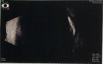
The retina exam of the left eye was unremarkable. The patient already had fluorescein angiography, ocular coherence tomography of the macula and a B – Scan examination done in another institution that showed results consistent with an osteoma.
She underwent three sessions of intravitreal anti – VEGF injection (Eylea) with monthly intervals. Post-treatment, she reported improvement in symptoms, visual acuity remains unchanged and repeat tests also showed better results.
The etiology of such tumor remains unknown, however, factors such as inflammation, trauma, hormonal state, calcium metabolism, environment, and heredity have been proposed.
Further research and discussion of these disease entities may help us further understand and ultimately treat the condition, in order to provide our patients with the best quality of life possible.
Ralph Anthony H. de Jesus
Asian Eye Institute
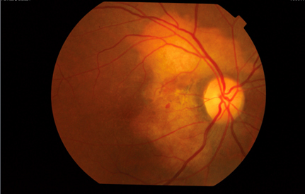
Fig. 1. Colored Photo, OD
Colored photo of the right eye showing a whitish lesion measuring approximately three-disc diameters in size and located temporal to the disc.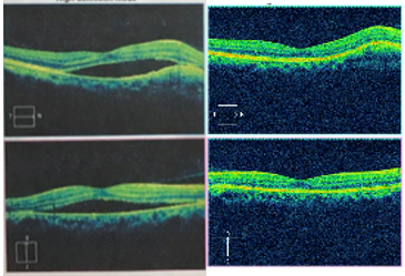
Fig. 2. Ocular Coherence Tomography Scan of the Macula, OD
Macular OCT Scan of the right eye pre-treatment showing the presence of a subretinal fluid in the area of the lesion. Post-treatment, there is resolution of the previously noted subretinal fluid.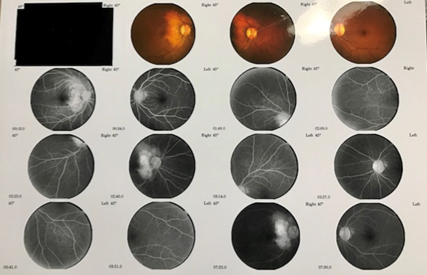
Fig. 3. Fluorescein Angiogram, OU
FA shows a hyperfluorescent lesion that did not increase in size or intensity towards the late phase in the right eye. The left eye is unremarkable.